Digital Pathology – Transforming the Future of Lab Testing
Traditional Pathology Scenario
A typical pathology workflow is a complex series of events, including a manual review of glass slides that ultimately results in diagnosis. However, the process of analyzing stains on a glass slide is lengthy and complicated. When a patient is referred to a pathologist, a sequence of actions is initiated, starting from extracting the tissue from the patient to offering an appropriate diagnosis. A bio-engineer prepares a sample of the extracted tissue for microscopy and then passes on the prepared slide to a pathologist, who specializes in cell and tissue diagnostics. If the pathologist finds difficulty in judgment, he/she consults with colleagues who can be in a different lab, state, or country altogether. The sample needs to be packaged, labeled, and mailed to the intended lab for remote consultation. The package also requires to be registered and delivered to the correct pathologist. However, the complete procedure can be quite expensive due to the cost incurred on packaging and courier services. This whole process might take several days to weeks and might cause damage to slides during transit. It also becomes difficult for the pathologist to search for old slides, images, and case information in the archives of hospitals. In addition, the results depend on the specialty and not on the personal judgment or mental state of the pathologist, as it may lead to lowering the level of accuracy.
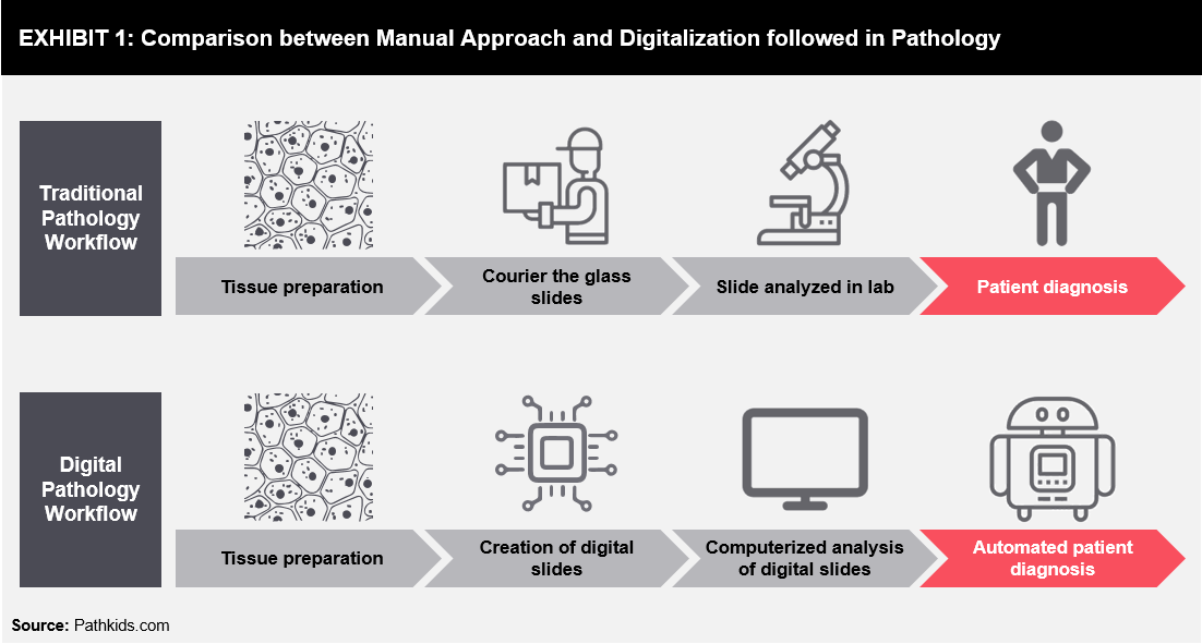
Digital pathology has been introduced to overcome numerous limitations associated with traditional pathology. Digital pathology is a computerized, image-based (digital slides) system for managing and interpreting information. The digital slide is a complete representation of a glass microscope slide, which can be viewed at any magnification, including intermediate magnifications not available on standard microscopes. The slides can be accessed remotely anywhere in the world for delivering consistent, rapid, and accurate results, as compared to traditional methods. Workflow difference in traditional and digital pathology is shown in Exhibit 1.
The pathological practice relies on the traditional approach, thus creating a challenging scenario for pathologists to deliver accurate results on time. Longer wait-time duration causes inconvenience to patients and further leads to a delay in treatment. Some of the most significant challenges are highlighted below:
- Slide storage: Most pathology labs (both individual and hospital-linked) lack a proper storage facility for the storage of slides. Many of these entities either do not have any plans or funds for expansion of the facility. The increasing demand for healthcare has contributed to the growth of the slide volume, which is increasing drastically year-on-year. With this increased volume, it becomes even more challenging to navigate and retrieve the slides, if required. There are higher chances of slides getting lost or deteriorated over time if the storage is not appropriate.
- Limited specialist’s opinion: Pathologists and histotechnologists are both a scarce resource in both hospitals and pathology labs. Hospitals and labs in remote locations may not have access to a specialist, as a result of which, either the patient has to travel to the referred specialist or samples have to be sent. In both cases, the process becomes lengthy, complicated, and time-consuming. Additionally, in urban areas, the emergence of new concepts such as laboratory consolidation for small-scale hospitals or ambulatory surgery centers leaves no room for a specialist to be present on-site.

- Pathologists are scarce assets: Pathologists are limited assets worldwide, and their number is predicted to decrease eventually. With a shortage in the number of pathologists in countries such as the United States, it is predicted that practicing pathologists will begin to retire at an alarming rate, peaking in 2021 faster than the current replacement rate. In some parts of Africa, there is only one pathologist present for every 1.5 million people. According to the Chinese Pathologist Association, there are only 20,000 licensed pathologists in China, with a population of over 1.4 billion people.
Digital workflow in pathology has the potential to address all these challenges.
Era of Digitalization in Pathology
With the introduction of digitalization in pathology, difficulties and barriers faced by the pathologists and patients due to traditional pathology workflow can be quickly addressed. The concept involves the acquisition, management, sharing, and interpretation of the pathology information in a digital manner.
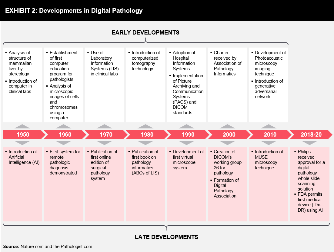
Digital pathology is not a new concept, and its history goes back around 100 years when specialized equipment was first introduced, and images from a microscope were captured onto a photographic plate via a camera. Telepathology emerged around 50 years back; however, over the past decade, pathology has undergone a real digital transformation, gradually shifting from analog to an electronic environment. The timeline of digitalization in pathology has been represented in Exhibit 2.
The digital pathology system is based on four key components:
- Capturing images (of slides) using digital scanners: Glass slides are represented in the digital form by using whole slide scanning, along with proper tools in an indicative manner. High-resolution scanning is performed, along with adequate color depth, so that images can be reproduced with the necessary magnifications required for diagnostic and research applications.
- Storing and archiving the images: The strategy of storing virtual slides is mainly dependent on the intended use. Local storage is sufficient for local diagnosis, while in certain instances, off-site storage may be required for remote consultation, using different options, such as Redundant Array of Independent Disks (RAID), optical/tape-based, cloud storage, or a combination approach.
- Editing and modifying captured images: Digital slides provide the opportunity to modify images according to the pathologist’s requirement, such as magnification, zoom-in/out, etc.
- Viewing and sharing images: Digital slides can be easily shared with different stakeholders, such as doctors, patients, research institutes, etc. Some software also support advanced visualization tools, enabling users to view multiple images in a single frame. Whole Slide Imaging (WSI) supports clinical diagnostics and image viewing in association with the patient’s clinical history, along with other slides or images that may have been acquired from the same patient (e.g., serial sections, IHC, grossing photos, radiology, etc.).
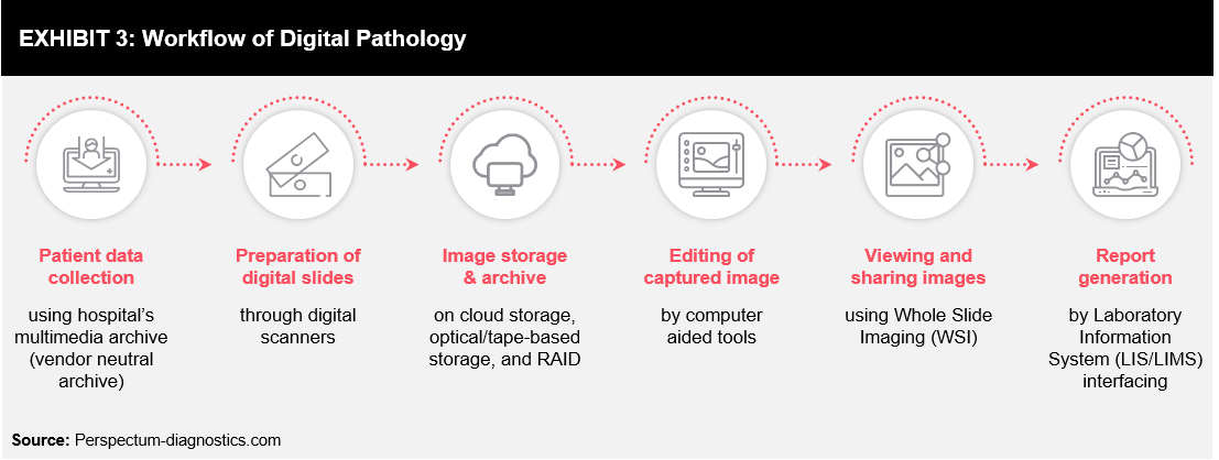
Whole Slide Imaging (WSI) technology, also known as “virtual or wide-field microscopy,” provides high-resolution digital images and relatively high-speed digitalization of glass slides of different samples. This makes the digitalization of sample slides more beneficial, interactive, and easy-to-share. The rapid progress of WSI technology, along with advances in software applications, Laboratory Information System (LIS/LIMS) interfacing and high-speed networking have made it possible to integrate digital pathology into pathology workflows. An illustrative workflow of digital pathology using WSI technology is explained in Exhibit 3.
Benefits and Limitations of Digital Pathology
Digital pathology is rapidly gaining momentum as a proven and essential technology, with specific support for education, tissue-based research, drug development, and the practice of human pathology throughout the world. It is an innovation committed to reduce laboratory expenses, improve operational efficiency, enhance productivity, and improve treatment decisions as well as patient care. Digital pathology comes with various benefits; some of these are listed below:
- Improved analysis: Algorithms provided for analyzing slides are objective, accurate, and faster than microscopy. With rapid access to prior cases, long-term predictive analytics is possible for greater accuracy.
- Reduced errors: Digital slides eliminate the risk of breakage of the glass slides, and barcoding leads to a reduction in the risk of misidentification.
- Better visualization: Digitalization enables live zooming and multiple angle views, resulting in better analysis. It provides pathologists with annotation of slides and dashboard views of data and annotations.
- Improved workflow: Central storage enables a streamlined workflow and better outsourcing. It also allows flexible work schedules and secure remote access.
- Reduced turnaround time: Reduction in the time required for slide retrieving, data matching, and organizing results has significantly reduced the turnaround time compared to manual reviews.
- Cost-effective: It has reduced costs by eliminating multiple steps and transportation services.
- Breaking geographical barriers: Digitalization has brought experts closer to the virtual world. It also provides an opportunity for teaching, training, and sharing expertise in remote locations.
Although there are various benefits, only limited providers have adopted digitalization in the pathology workflow for primary diagnosis due to various limitations:
- Data storage: Large data storage capacity is required for high-resolution images, which serves to be a challenge, along with their transmission through systems.
- Non-standardization: With no standardization in digital pathology processes, there are limited choices of solutions currently available in the market.
- High investments: Significant investments are required to set-up or upgrade to digital pathology networks. The ambiguity of potential return on investment is slowing the adoption and growth rate for digital pathology.
- Data safety: Robust networks are required to process data and ensure high-level data confidentiality, as per regulatory guidelines.
Trends in Digital Pathology
Established biopharmaceutical companies and top clinical research organizations have adopted the concept of digital pathology to streamline their drug development processes. The distinct opportunity exists for the potential use of digital pathology in the analysis of emerging companion diagnostics and novel theragnostics. Digital pathology can be relevant with the advent of assays such as markers or multiplex, which are difficult to discern with the human eye. Digital pathology has started to expand and will be facilitating advanced diagnostic skills for pathologists. Well-established digital pathology companies need to consider building the required business framework to support the development of precision medicine using digital pathology.
Some of the companies, such as Inspirata Inc. and MD Biosciences, have adopted different approaches to increase their foothold in the digital pathology domain. Inspirata offers “Digital Informatics Solution” that streamlines case management and communication among the entire care team, whereas MD Biosciences offers cutting-edge digital pathology imaging equipment for rapid, high-resolution digitalization of whole slides for reviewing and assessing through their secure and cloud-based image management system.
There are several algorithms being developed (e.g., pattern recognition algorithms) that improves accuracy, reliability, specificity, and productivity. Computer-assisted Image Analysis (CAIA) has been used to score certain immunohistochemical stains, although this gives all pathologists the same yardstick for scoring immunohistochemistry findings in cancer.
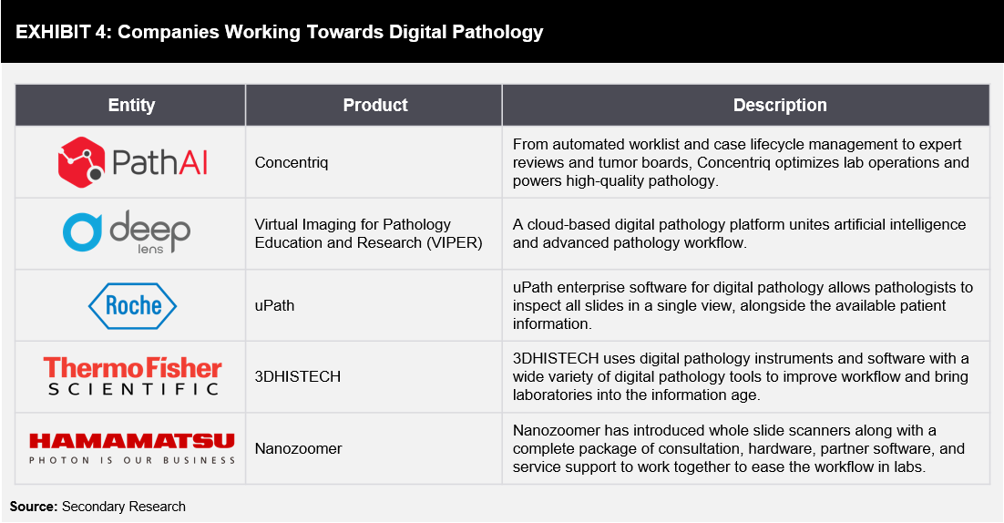
With the increased use of exponential technologies such as Artificial Intelligence (AI) and Machine Learning (ML) in digital pathology, enhanced translational research, Computer-aided Diagnosis (CAD), and personalized medicine are expected to grow in the near future. Exhibit 4 lists some of the companies actively working in digitalizing pathology.
Conclusion
Digital pathology is a disruptive technology that has encouraged the practice of virtual pathology and has the potential to replace traditional pathology practices. Digitalization and automation of the whole process from image capture, analysis, and clinical decision support, have reduced the lengthy procedures, the time required for analysis, and manual intervention of pathologists. With the advent of digital pathology, pathologists can interact with each other for remote consultations and provide an accurate analysis. To date, digital pathology is mainly supporting pathologists for secondary diagnosis and validation of analysis. It is assumed that digital pathology is not meant for taking pathologists out of the picture, but to support them for rapid and accurate decision-making. Key stakeholders in the healthcare industry are investing and working towards digitalizing pathology using artificial intelligence, cloud computing, and other advanced analytics tools. With the emerging visualization and data analytics tools, digital pathology will undoubtedly allow pathologists to make a more accurate and consistent diagnosis in the near future. However, it seems that there is a long and tough path ahead for proving the efficacy of digital pathology tools and pass regulatory hurdles. A variety of digital solutions are already available in the market for migrating the entire workflow of pathologists from manual to digital; these solutions have helped overcome barriers and address associated challenges with the traditional pathology workflow.
References
- https://www.pathkids.com/digital.htm
- http://www.clpmag.com/2017/10/digital-pathology-gives-rise-computational-pathology/
- https://insights.samsung.com/2018/05/17/the-digital-future-of-pathology/
- https://www.nature.com/articles/s41571-019-0252-y
- https://thepathologist.com/outside-the-lab/facing-the-digital-future-of-pathology
- https://perspectum-diagnostics.com/products-and-research/digital-pathology
- https://www.pathai.com/
- https://www.deeplens.ai/
- https://diagnostics.roche.com/global/en/products/instruments/upath-enterprise-software.html
- https://www.3dhistech.com/
- https://nanozoomer.hamamatsu.com/us/en/index.html
- https://www.ncbi.nlm.nih.gov/pmc/articles/PMC3074674/
- https://healthcare-in-europe.com/en/news/the-challenges-of-digital-pathology.html
- https://medcitynews.com/2016/11/digital-pathology-challenges/
- https://www.ncbi.nlm.nih.gov/pmc/articles/PMC2941968/
- https://www.philips.co.uk/healthcare/sites/pathology/about/what-is-digital-pathology
- https://www.leicabiosystems.com/knowledge-pathway/digital-pathology/
- https://medcitynews.com/2019/04/ai-pathology-startup-60m/?rf=1
- https://academic.oup.com/labmed/article-pdf/38/6/341/24960144/labmed38-0341.pdf


